
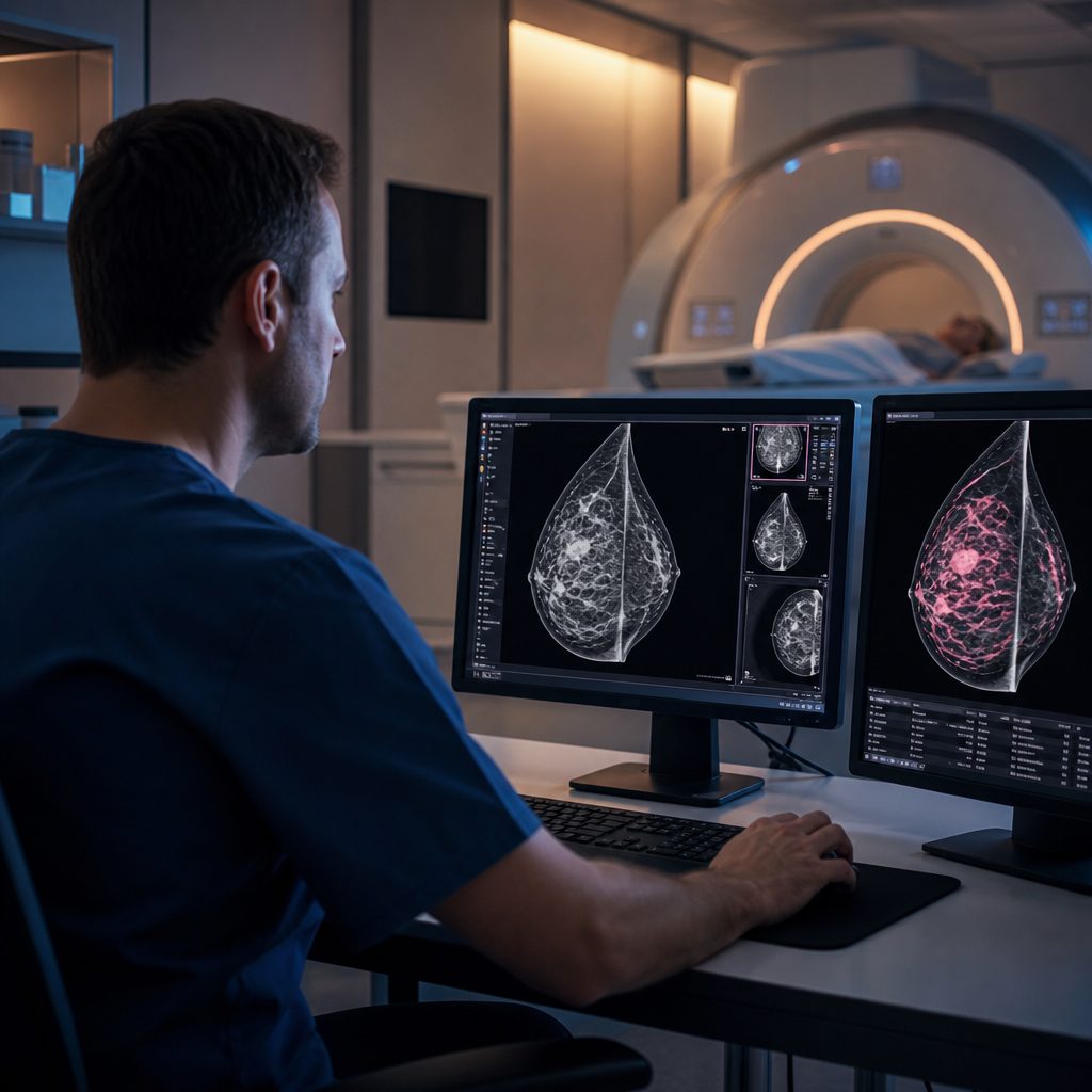
Advanced Imaging Techniques That Improve Breast Cancer Detection in High-Risk Women
Women with elevated breast cancer risk face a significant challenge: standard mammography alone may not catch aggressive cancers early enough. Dense breast tissue, genetic mutations such as BRCA1 and BRCA2, and a family history of the disease all increase the likelihood that routine screening will miss lesions in their earliest, most treatable stages. For these women, supplemental imaging methods have become essential tools in the fight for early detection.
Understanding which advanced imaging options exist, when they are appropriate, and how they complement traditional screening can make the difference between catching cancer at stage one versus stage three. This article examines the imaging techniques that extend beyond mammography and the evidence supporting their use in high-risk populations.
Who Qualifies as High Risk
Risk stratification determines which women benefit from enhanced surveillance. The baseline definition includes those with a lifetime breast cancer risk of 20 percent or higher, calculated using models such as the Tyrer-Cuzick or BRCAPRO tools. Women carrying pathogenic variants in BRCA1, BRCA2, TP53, PTEN, or other high-penetrance genes fall into this category automatically.
Personal history matters as well. Prior chest radiation between ages 10 and 30, typically for Hodgkin lymphoma, creates lasting tissue changes that elevate cancer risk decades later. Dense breast tissue, classified as heterogeneously dense or extremely dense on mammography reports, reduces the sensitivity of X-ray imaging and itself confers a modest increase in cancer incidence. First-degree relatives diagnosed before menopause or with bilateral disease also shift risk calculations upward.
Supplemental Screening Options
Breast Ultrasound
Whole-breast ultrasound detects cancers mammography misses in dense tissue, adding approximately three additional cancers per 1,000 women screened. The technique uses sound waves rather than radiation, making it safe for repeated use. Radiologists look for irregular masses with shadowing, microcalcifications, and vascularity patterns that suggest malignancy.
The trade-off involves false positives. Ultrasound identifies many benign findings—fibroadenomas, cysts, areas of fibrocystic change—that require follow-up imaging or biopsy to exclude cancer. Studies show callback rates increase when ultrasound supplements mammography, which can create anxiety and additional healthcare visits. Despite this, the method remains valuable for women whose breast density would otherwise obscure small tumors.
Magnetic Resonance Imaging
Breast MRI stands as the most sensitive imaging tool available for high-risk screening, detecting 90 percent or more of cancers compared to mammography's 40 to 60 percent in dense breasts. The examination uses powerful magnets and contrast agents to create detailed cross-sectional images. Radiologists analyze enhancement patterns, with rapid uptake and washout of contrast suggesting malignancy. For women with genetic mutations or a calculated lifetime risk above 20 percent, annual mri alongside mammography has become the standard of care, supported by guidelines from the American Cancer Society and other professional bodies.
Cost and accessibility limit MRI for lower-risk populations. Each scan requires 30 to 45 minutes in the machine, intravenous contrast administration, and specialized radiologist interpretation. False positives remain common, with benign enhancement from hormonal changes, fibroadenomas, or papillomas prompting further workup. Insurance coverage varies, typically approving MRI only when documented risk factors meet specific thresholds.
Contrast-Enhanced Mammography
This newer technique injects iodinated contrast before acquiring low- and high-energy mammogram images. Subtraction processing highlights areas of abnormal blood flow, similar in concept to MRI but performed on standard mammography equipment. Early studies suggest sensitivity approaching that of MRI at roughly one-third the cost and without the claustrophobia or lengthy exam time.
Contrast-enhanced mammography has not yet accumulated the decades of outcome data supporting MRI. Questions remain about optimal recall thresholds, interpretation standards, and long-term cancer detection rates. Still, the method offers promise for centers lacking MRI capacity or for patients with contraindications to magnetic imaging, such as certain implanted devices.
Timing and Frequency Considerations
Screening schedules for high-risk women differ from average-risk protocols. Guidelines recommend starting annual mammography and MRI at age 30 for BRCA carriers, or eight years after chest radiation exposure. Some clinicians stagger the two modalities six months apart to achieve semi-annual surveillance, though evidence supporting this approach over concurrent annual screening remains limited.
Menstrual cycle timing affects MRI interpretation. Background parenchymal enhancement peaks during the luteal phase, creating false-positive findings that can obscure or mimic tumors. Scheduling MRI during days 7 through 14 of the cycle, when estrogen levels are moderate and progesterone remains low, reduces unnecessary callbacks. Postmenopausal women face no such timing constraints, and those interested in comprehensive health maintenance beyond breast imaging may benefit from understanding broader preventive care approaches for their life stage.
Evaluating Detection Performance
Sensitivity and specificity measure how well imaging identifies true cancers while avoiding false alarms. Mammography sensitivity drops from 85 percent in fatty breasts to below 50 percent in extremely dense tissue. Adding ultrasound raises sensitivity modestly but increases false positives substantially. MRI achieves the highest sensitivity, often exceeding 95 percent, at the cost of lower specificity—around 85 percent—meaning one in seven findings flagged on MRI proves benign after biopsy.
Detection is only part of the equation. Interval cancers, those diagnosed between screening rounds, occur more frequently in high-risk women despite intensive surveillance. These tumors often display aggressive biology, growing rapidly between annual exams. Research into shorter screening intervals or novel biomarkers continues, aiming to catch these fast-growing cancers earlier. Organizations such as Scripps Health have compiled guidance on when different screening methods become appropriate based on age and risk factors.
Managing the Emotional Impact of Intensive Screening
High-risk surveillance programs generate anxiety. Frequent imaging appointments, callback letters, and biopsies create a cycle of anticipation and relief—or dread. Studies document elevated distress levels in women undergoing MRI screening, particularly during the waiting period between imaging and results.
Support systems help. Many cancer centers offer genetic counseling, psychological services, and peer support groups specifically for high-risk women. Understanding that most callbacks resolve as benign findings—often simple cysts or fibroadenomas—provides perspective, though it rarely eliminates worry entirely. Some women find comfort in the knowledge that intensive screening detects cancers at the earliest possible stage, when treatment options are broadest and outcomes most favorable.
Cost and Access Challenges
Insurance coverage for supplemental imaging depends on documented risk factors. Most US insurers cover annual breast MRI for BRCA carriers and others meeting high-risk criteria, but prior authorization requirements and out-of-pocket costs vary widely. Women with dense breasts but no other risk factors often face coverage denials for MRI, leaving ultrasound as the only supplemental option.
Geographic disparities compound financial barriers. Rural areas and underserved communities frequently lack access to breast MRI, requiring travel to regional medical centers. Mobile MRI units and telemedicine interpretation have begun addressing these gaps, but availability remains inconsistent. Advocacy for insurance coverage of supplemental screening continues at state and federal levels, with several US states now mandating coverage for women with dense breasts.
The Path Forward
Advanced imaging technologies continue to evolve. Abbreviated MRI protocols reduce scan time to less than 10 minutes while maintaining detection rates, potentially improving access and reducing cost. Artificial intelligence algorithms now assist radiologists in identifying suspicious findings, with some systems matching or exceeding human performance in research settings. Three-dimensional mammography, or tomosynthesis, has become standard in many centers, offering improved visualization of overlapping tissue structures.
For high-risk women, the question is no longer whether to pursue enhanced screening but which combination of methods best balances cancer detection against false positives, cost, and convenience. Shared decision-making with clinicians who understand individual risk profiles, imaging performance characteristics, and patient preferences remains essential. Early detection saves lives, and for those at elevated risk, the tools to achieve it have never been more powerful.